Structure of the recombinant N-terminal lobe of human lactoferrin at 2.0 A resolution.
Day, C.L., Anderson, B.F., Tweedie, J.W., Baker, E.N.(1993) J Mol Biol 232: 1084-1100
- PubMed: 8371268
- DOI: https://doi.org/10.1006/jmbi.1993.1462
- Primary Citation of Related Structures:
1LCT - PubMed Abstract:
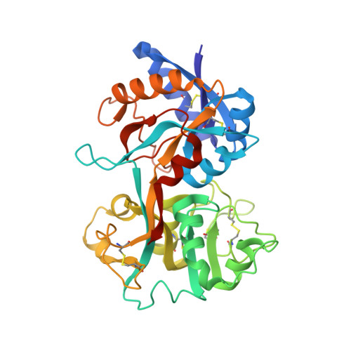
The three-dimensional structure of the N-terminal half-molecule of human lactoferrin, LfN, prepared by recombinant DNA methods, has been determined by X-ray crystallography at 2.0 A resolution. The protein is in its iron-bound form and is deglycosylated. X-ray diffraction data were obtained by diffractometry to 3.2 A resolution and synchrotron data collection, using Weissenberg photography with imaging plates, to 1.8 A resolution. The structure was solved by molecular replacement, using the N-lobe of native diferric human lactoferrin (Lf) as search model. Restrained least squares refinement (program TNT) has resulted in a model structure with an R-factor of 0.184 for all data 34,180 (reflections) in the resolution range 8.0 to 2.0 A. The model comprises 2490 protein atoms (residues 4 to 327), 1 Fe3+, 1 CO3(2-) and 180 solvent molecules, all regarded as water. The structure of LfN is essentially the same as that of the N-lobe of intact Lf, being folded into two similar alpha/beta domains, with the Fe3+ and CO3(2-) bound in a specific site in the interdomain cleft. These details are not affected by either deglycosylation or expression in a non-native system. At the C terminus, however, the conformation of residues 321 to 333 is changed. Whereas in Lf residues 321 to 332 form a helix crossing between the domains at the back of the iron site, in LfN residues 321 to 326 have an extended conformation, forming a third interdomain beta-strand, and residues 328 to 333 appear disordered. The conformational change is attributed to the loss of stabilizing interactions from the C-lobe and is mediated by two Gly residues, at positions 321 and 323. It is further proposed that the conformational change is responsible for the more facile iron release properties of LfN, by its effect on the hinge mechanism and increased solvent exposure of residues near the back of the iron site. Other details of the polypeptide chain conformation and the binding site have also been analysed. Two cis-proline residues are found at positions 71 and 142. The bidentate binding of the CO3(2-) to the metal ion is unambiguous, and a network of hydrogen bonds in and around the binding site links the two domains. Clearly-defined amino-aromatic hydrogen bonds are found for Arg210, near the metal site, and some 31 internal water molecules have been identified, 15 of them in essentially discrete sites, and 16 in a cluster filling a cavity in the interdomain cleft.
Organizational Affiliation:
Department of Chemistry and Biochemistry, Massey University, Palmerston North, New Zealand.