1LFH

MOLECULAR REPLACEMENT SOLUTION OF THE STRUCTURE OF APOLACTOFERRIN, A PROTEIN DISPLAYING LARGE-SCALE CONFORMATIONAL CHANGE


Experimental Data Snapshot

  • Method: X-RAY DIFFRACTION
  • Resolution: 2.80 Å
  • R-Value Observed: 0.213 

wwPDB Validation   3D Report Full Report


This is version 1.3 of the entry. See complete history


Literature

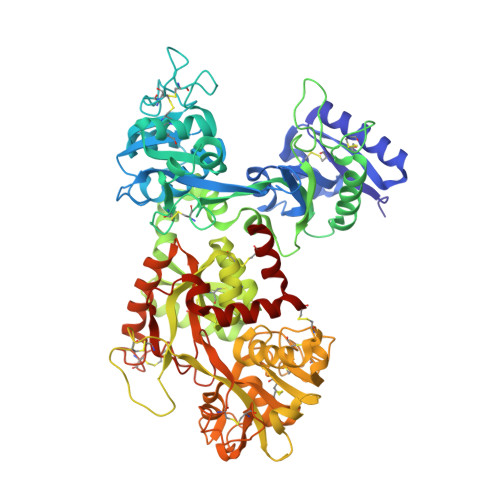
Molecular replacement solution of the structure of apolactoferrin, a protein displaying large-scale conformational change.

Norris, G.E.Anderson, B.F.Baker, E.N.

(1991) Acta Crystallogr B 47: 998-1004

  • DOI: https://doi.org/10.1107/s0108768191008418
  • Primary Citation of Related Structures:  
    1LFH

  • PubMed Abstract: 

    The crystal structure of an orthorhombic form of human apolactoferrin (ApoLf) has been determined from 2.8 A diffractometer data by molecular replacement methods. A variety of search models derived from the diferric lactoferrin structure (Fe2Lf) were used to obtain a consistent solution to the rotation function. An R-factor search gave the correct translational solution and the model was refined by rigid-body least-squares refinement (program CORELS). Only three of the four domains were located correctly by this procedure, however; the fourth was finally placed correctly by rotating it manually onto three strands of electron density which were recognized as part of its central beta-sheet. The final model, refined by restrained least-squares methods to an R factor of 0.214 for data in the resolution range 10.0 to 2.8 A, shows a large domain movement in the N-terminal half of the molecule (a 54 degree rotation of domain N2) and smaller domain movements elsewhere, when compared with Fe2Lf. A feature of the crystal structure is that although the ApoLf and Fe2Lf unit cells appear very similar, their crystal packing and molecular structures are quite different.


  • Organizational Affiliation

    Department of Chemistry and Biochemistry, Massey University, Palmerston North, New Zealand.


Macromolecules
Find similar proteins by:  (by identity cutoff)  |  3D Structure
Entity ID: 1
MoleculeChains Sequence LengthOrganismDetailsImage
LACTOFERRIN691Homo sapiensMutation(s): 0 
UniProt & NIH Common Fund Data Resources
Find proteins for P02788 (Homo sapiens)
Explore P02788 
Go to UniProtKB:  P02788
PHAROS:  P02788
GTEx:  ENSG00000012223 
Entity Groups  
Sequence Clusters30% Identity50% Identity70% Identity90% Identity95% Identity100% Identity
UniProt GroupP02788
Sequence Annotations
Expand
  • Reference Sequence
Small Molecules
Ligands 1 Unique
IDChains Name / Formula / InChI Key2D Diagram3D Interactions
CL
Query on CL

Download Ideal Coordinates CCD File 
B [auth A]CHLORIDE ION
Cl
VEXZGXHMUGYJMC-UHFFFAOYSA-M
Experimental Data & Validation

Experimental Data

  • Method: X-RAY DIFFRACTION
  • Resolution: 2.80 Å
  • R-Value Observed: 0.213 
  • Space Group: P 21 21 21
Unit Cell:
Length ( Å )Angle ( ˚ )
a = 152.09α = 90
b = 94.58β = 90
c = 55.79γ = 90
Software Package:
Software NamePurpose
TNTrefinement

Structure Validation

View Full Validation Report


Entry History 

Deposition Data

Revision History  (Full details and data files)

  • Version 1.0: 1993-10-31
    Type: Initial release
  • Version 1.1: 2008-03-03
    Changes: Version format compliance
  • Version 1.2: 2011-07-13
    Changes: Version format compliance
  • Version 1.3: 2017-11-29
    Changes: Derived calculations, Other