1POD

STRUCTURES OF FREE AND INHIBITED HUMAN SECRETORY PHOSPHOLIPASE A2 FROM INFLAMMATORY EXUDATE


Experimental Data Snapshot

  • Method: X-RAY DIFFRACTION
  • Resolution: 2.10 Å
  • R-Value Work: 0.193 
  • R-Value Observed: 0.193 

wwPDB Validation   3D Report Full Report


This is version 2.1 of the entry. See complete history


Literature

Structures of free and inhibited human secretory phospholipase A2 from inflammatory exudate.

Scott, D.L.White, S.P.Browning, J.L.Rosa, J.J.Gelb, M.H.Sigler, P.B.

(1991) Science 254: 1007-1010

  • DOI: https://doi.org/10.1126/science.1948070
  • Primary Citation of Related Structures:  
    1POD, 1POE

  • PubMed Abstract: 

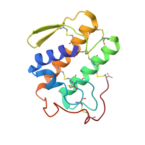
    Phospholipase A2 (PLA2) participates in a wide range of cellular processes including inflammation and transmembrane signaling. A human nonpancreatic secretory PLA2 (hnps-PLA2) has been identified that is found in high concentrations in the synovial fluid of patients with rheumatoid arthritis and in the plasma of patients with septic shock. This enzyme is secreted from certain cell types in response to the proinflammatory cytokines, tumor necrosis factor or interleukin-1. The crystal structures of the calcium-bound form of this enzyme have been determined at physiological pH both in the presence [2.1 angstrom (A) resolution] and absence (2.2 A resolution) of a transition-state analogue. Although the critical features that suggest the chemistry of catalysis are identical to those inferred from the crystal structures of other extracellular PLA2s, the shape of the hydrophobic channel of hnps-PLA2 is uniquely modulated by substrate binding.


  • Organizational Affiliation

    Department of Molecular Biophysics and Biochemistry, Yale University, New Haven, CT 06511.


Macromolecules
Find similar proteins by:  (by identity cutoff)  |  3D Structure
Entity ID: 1
MoleculeChains Sequence LengthOrganismDetailsImage
PHOSPHOLIPASE A2124Homo sapiensMutation(s): 0 
EC: 3.1.1.4
UniProt & NIH Common Fund Data Resources
Find proteins for P14555 (Homo sapiens)
Explore P14555 
Go to UniProtKB:  P14555
PHAROS:  P14555
GTEx:  ENSG00000188257 
Entity Groups  
Sequence Clusters30% Identity50% Identity70% Identity90% Identity95% Identity100% Identity
UniProt GroupP14555
Sequence Annotations
Expand
  • Reference Sequence
Small Molecules
Ligands 1 Unique
IDChains Name / Formula / InChI Key2D Diagram3D Interactions
CA
Query on CA

Download Ideal Coordinates CCD File 
B [auth A]CALCIUM ION
Ca
BHPQYMZQTOCNFJ-UHFFFAOYSA-N
Experimental Data & Validation

Experimental Data

  • Method: X-RAY DIFFRACTION
  • Resolution: 2.10 Å
  • R-Value Work: 0.193 
  • R-Value Observed: 0.193 
  • Space Group: P 61 2 2
Unit Cell:
Length ( Å )Angle ( ˚ )
a = 76.3α = 90
b = 76.3β = 90
c = 90.6γ = 120
Software Package:
Software NamePurpose
X-PLORmodel building
PROFFTrefinement
X-PLORrefinement
X-PLORphasing

Structure Validation

View Full Validation Report


Entry History 

Deposition Data

Revision History  (Full details and data files)

  • Version 1.0: 1993-10-31
    Type: Initial release
  • Version 1.1: 2008-03-03
    Changes: Version format compliance
  • Version 1.2: 2011-07-13
    Changes: Version format compliance
  • Version 1.3: 2011-11-16
    Changes: Atomic model
  • Version 2.0: 2019-07-17
    Changes: Atomic model, Data collection, Other, Refinement description
  • Version 2.1: 2019-08-14
    Changes: Advisory, Data collection, Derived calculations, Refinement description